#20. Eye Health NGO
This is probably one of the commonest images you’ll find in this list, but there’s an interesting backstory behind it. Lying on the floor, a man is receiving an eye exam in a makeshift clinic founded by the famous NGO, Unite For Sight.


This NGO aims to improve global eye health, and over the past years, it has provided over 90,000 cataract surgeries and has assisted nearly 2,5 million people. This picture aims to raise awareness on the issue of eye health.
#19. Digital Illustration With Electric Pen
This image may look like a painting, but it’s actually a digital illustration created using an electric pen. The image represents Rita Levi-Montalcini, the woman who won the Nobel Prize in Physiology in 1986 for discovering nerve growth factor.

For those of you unfamiliar with the art world, digital or computer illustration is the use of digital tools to produce illustrations through the manipulation of the artist. A digital pen, in turn, is an input device which converts handwritten drawings or information into digital data. Crazy, right?
#18. Mouse’s Placentas Through Confocal Microscopy
If we let our imagination carry us away, the shapes that appear in the picture look like fossilized mandarine segments. But, believe it or not, what you see below is the developmental stages of mouse placentas. Yikes!

This image was obtained using confocal microscopy, also known as confocal laser scanning microscopy, which is an optical imaging technique meant to increase the optical resolution of an image. You’re also probably wondering how these contrasting colors and shades were achieved, and here’s the answer. The placentas were stained to show three different proteins: the blue represents the nucleus, the green shows the presence of trophoblasts, while the red indicates blood vessels.
#17. Scanning Electron Microscopy
This purple figure was obtained using a scanning electron microscope, a type of microscope that creates images of a sample by scanning the surface with a focused beam of electrons. But can you guess what is represented in this image?

What you see in the image are actually microRNAs, a small RNA molecule found in plants, animals, and certain viruses. These molecules control the growth and functioning of cells and scientists are investigating whether or not they can be used as a possible cancer therapy.
#16. Intraocular Lenses
The technological advances in the photography world allow photographers to take pictures with much greater detail. This one here is a picture of an eye, but if you look closely, there’s a thin transparent object covering it. Can you guess what it is?
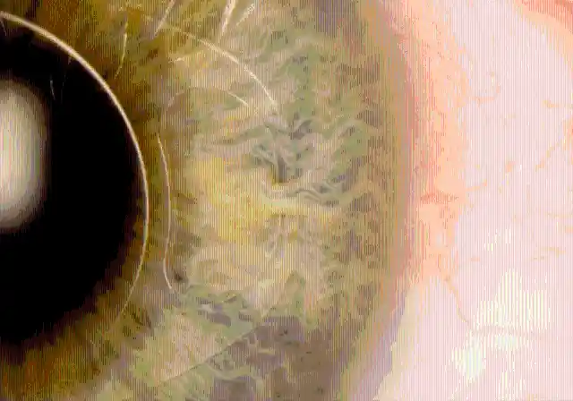

This object happens to be an intraocular lens, also known as iris clip, which is used to treat diseases such as cataracts and nearsightedness. Placing intraocular lenses in your eyes requires surgery. The patient whose eye is portrayed in the picture regained full vision after undergoing this surgery. Impressive, right?
#15. Pig Eye Model Through 3D Printing
The image below was created through 3D printing, a technique that consists in building a three-dimensional object from a computer-aided design model. This is usually carried out by adding material layer by layer. But what is this 3D object supposed to be?

This white object is the model of a pig’s eye. The indent you can see to the right is the pupil, which allows light into the eye. The left side represents the blood vessels in the muscles that surround the iris.
#14. Bones Through Computer-Generated Imagery
This picture, taken by the artist Oliver Burston, is part of a series named Stickman – The Vicissitudes of Crohn’s. The images that belong to this series are based around the character Stickman, an alter ego of the artist, who happens to suffer from Chron’s disease.
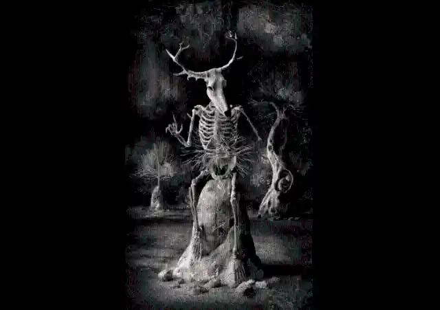
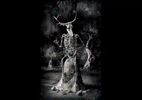
This picture, which was created through computer-generated imagery, is supposed to represent the symptoms generated by Chron’s disease, a chronic gastroenterological condition which produces weight loss and bone fragility. The picture is creepy, but at the same time impressive.
#13. 3D Model Of A Human Brain
If you take a glance at the image below, it kind of looks like an item made out of some sort of wool, right? A small pashmina, for instance. Well, if you had the same impression as I did, then I’m afraid to tell you that you couldn’t be wronger. This image is actually a 3D model of a human brain. But how was it created?
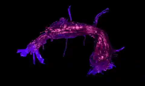
This image was made with a scan of a human brain using a 3D modeling technique called tractography. Tractographies are used to visually represent certain parts of the brain such as nerve tracts. What you see in this picture is actually white matter neural pathways which connect the two regions of the human brain responsible for language.
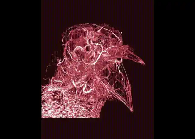
#12. Computed Tomography Scan Of A Parrot
This weird and scary image seems like a painting, but it was created using extremely sophisticated procedures. What you see below is the combination of 2,933 images, each one 0.1 millimeters thick. But how was the final product achieved?

These nearly 3,000 images were taken with Computed Tomography scans, a device that uses computer-processed combinations of X-rays to produce tomographic images of certain areas of the body. In this case, the specific combination of X-rays is meant to represent the parrot’s blood vessels and bones. However, this model was slightly manipulated with digital imaging software.
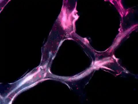
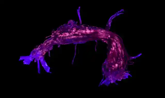
#11. Synthetic Gel Through Confocal Microscopy
I don’t know about you, but I think this would make a great painting in a contemporary art museum. Look at how striking those shades of violet are! If you’re not a science expert, it’s hard to tell what this image represents. Does anyone dare guess?
This image was also created by the use of confocal microscopy, a technique that has already been explained in previous slides. In this case, the image shows the growth of neural stem cells on a synthetic gel called PEG (polyethylene glycol).
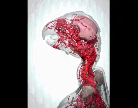
#10. Computed Tomography Scan Of A Parrot
This image seems like a digitally animated cartoon, doesn’t it? Well, actually, its a computed tomography scan of a gray parrot. Computed tomography scans, also known as CT scans, use x-rays to show the body’s bones and blood vessels.
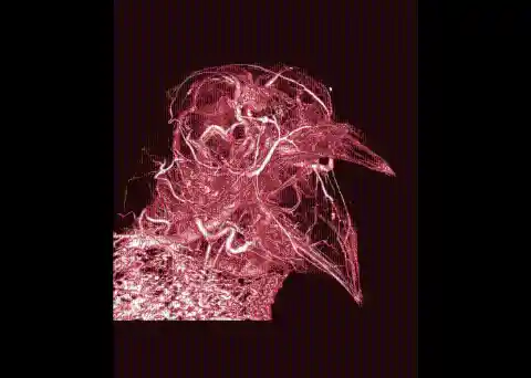
Therefore, most of what you see in this picture is the parrot’s blood vessels. The dense mass of blood that you can see below its neck helps the parrot regulate its body temperature through a process known as thermoregulation.
#9. Digital Art Of A Cell Membrane
This image was created using digital art. The silver ring-like shapes are channels spanning the membrane of a human cell. The light blue spheres, in turn, may look like bubblegum, but they represent cargo moving through the cell.
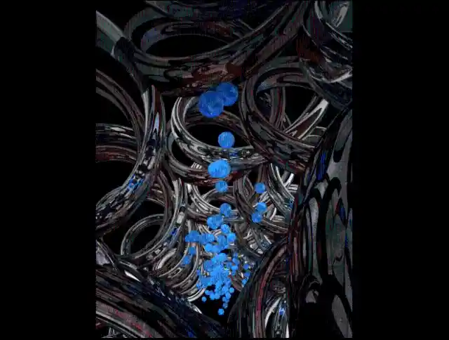

These models were created using a 3D modeling and animation program called Strata Design 3D CX. They were then saved as PSD files and finally colorized using Photoshop. They did a great job adding the colors!
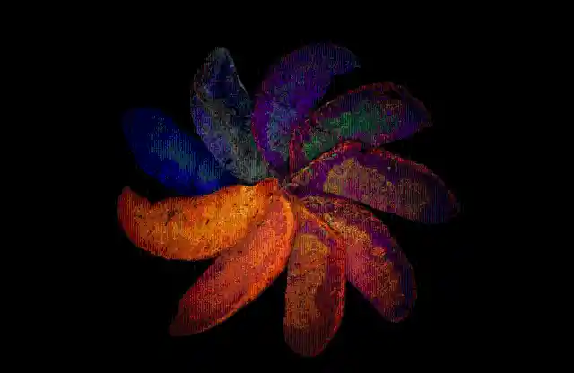
#8. Mouse’s Retina Through Confocal Microscopy
This is one of the many images in this list that have been created with the confocal microscopy technique. This leaf-like image represents the retina of a mouse. Retinas are at the very back of the eye and they are responsible for transforming light into nerve signals.
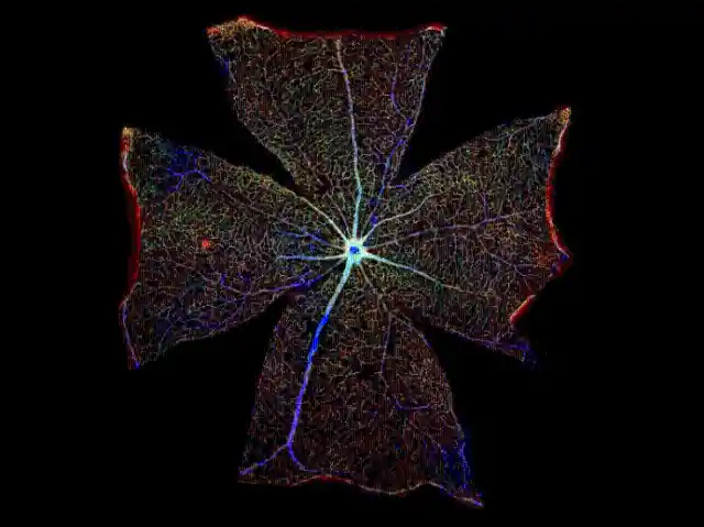
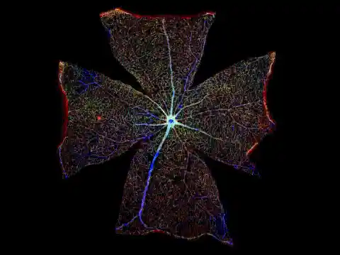
If you’re interested in greater detail, what you see isn’t the entire retina, but just a single cell called astrocyte. Astrocytes are star-shaped cells found in the brain-retinal barrier.
#7. Graphical Visualization Of Twitter Data
Created by Eric Clarke, Richard Arnett, and Jane Burns, this illustration is one of the strangest ones included in this list. This image is a graphical visualization of data extracted from tweets that used the hashtag #breastcancer.
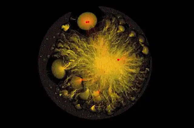

The Twitter users who used this hashtag are represented by small yellow dots called nodes, and the lines connecting the nodes symbolize the conversations that the Twitter users had. The sizes of the dots vary according to each user’s amount of connections and online presence. How many days do you think it took them to make this?
#6. Photomacrography Of A Hawaiian Bobtail Squid
The creature that you can see below is a Hawaiian bobtail squid. If you ask me, it looks like a computerized image or a digitally manipulated picture, but surprisingly, it’s neither of the above. Can you guess how this picture was created?
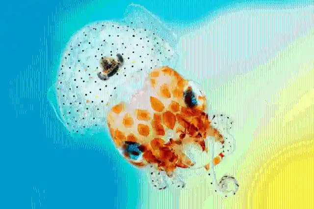

This squid was photographed using a complex and sophisticated technique known as photomacrography. This procedure uses specialized lenses as well as multiple stitched images to create a highly detailed picture. If you come across a squid in real life, there’s no way you’ll be able to spot its yellow and black dots or the different shades of orange it has in its head!
#5. Mouse’s Spine Through Confocal Microscopy
I know what you’re thinking: these three images – which, to be honest, kind of look like winter slippers, don’t they? – are completely surreal. But believe it or not, they’re real-life images of the developing spinal cord of an embryonic mouse. Who would’ve thought?
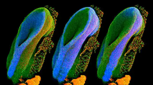

But this no ordinary image, as it was made employing a complex technique known as confocal microscopy. As we have seen, through this technique, lights such as lasers are stacked to create a 3D reconstruction of a certain microscopic image. Cool, huh?
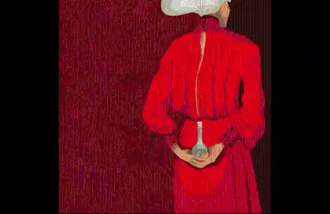
#4. “Hidden Learning” Painting
This may seem like a digitally manipulated image, but it’s actually the only regular painting on this list. It was made by illustrator Sophie McKay Knight and titled Hidden Learning. The painting is part of the so-called Chrysalis project, an artistic movement that seeks to bring together female scientists.


This painting is meant to represent the things that women feel they keep hidden in the work environment, such as the constant struggle between their career and home life. The woman is carrying a sort of veil, and only professional scientists will have noticed that it’s made up of the molecular structure of a sugar molecule,
#3. Cat Skin Through Polarized Light Microscopy
I know what you’re thinking: this has got to be a painting! Believe it or not, this image shows the different types of tissue found in some sections of cat skin. But how was this image created? Why all those colors?

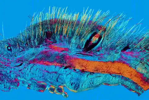
This cat skin image was produced with an optical microscopy technique called polarized light microscopy. This technique involves the illumination of a sample with polarized light. In other words, this form of microscopy splits light traveling through different materials and directions, consequently creating a wide range of colors.
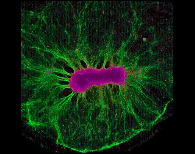
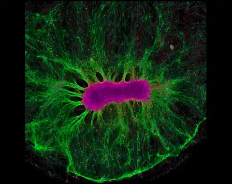
#2. Zebrafish Embryo Trough Confocal Microscopy
This crazy image full of fluorescent lights looks like a digitally manipulated painting, but it was actually created using confocal microscopy. You’ll never guess what this image represents, as it is supposed to be a zebrafish embryo.
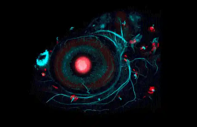
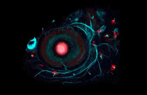
But how can we explain such colors? The embryo has been injected with a dyed red gene which researchers hope to manipulate. The scientists are interested in observing whether the injection of this gene will affect the fish’s schooling behaviors and predator responses.
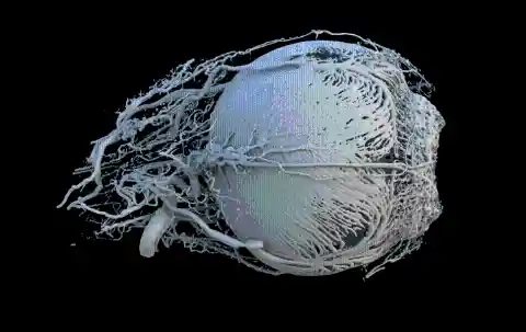
#1. Super-Resolution Image Of A Nucleus
It’s hard to believe that this is anything but a painting, right? Well, believe it or not, this image was obtained using a technique called super-resolution imaging. This procedure enhances the resolution of an imaging system and is frequently used in super-resolution microscopy.
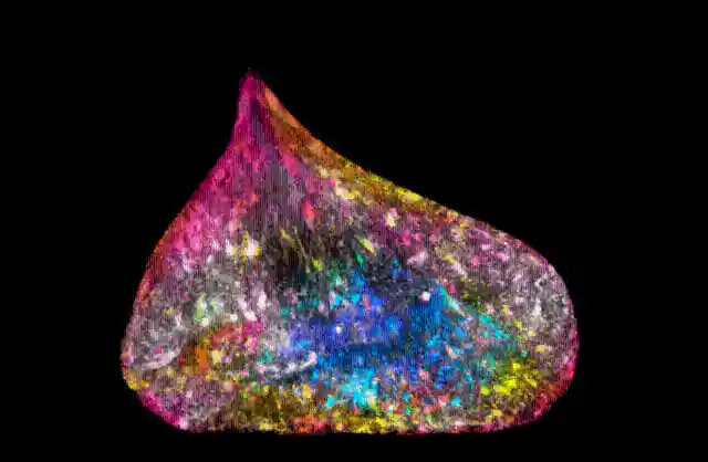

This super-resolution image shows the nucleus of a DNA cell of a human lung. To be more precise, it’s one of two cells that have just been divided through a process of mitosis. Impressive!